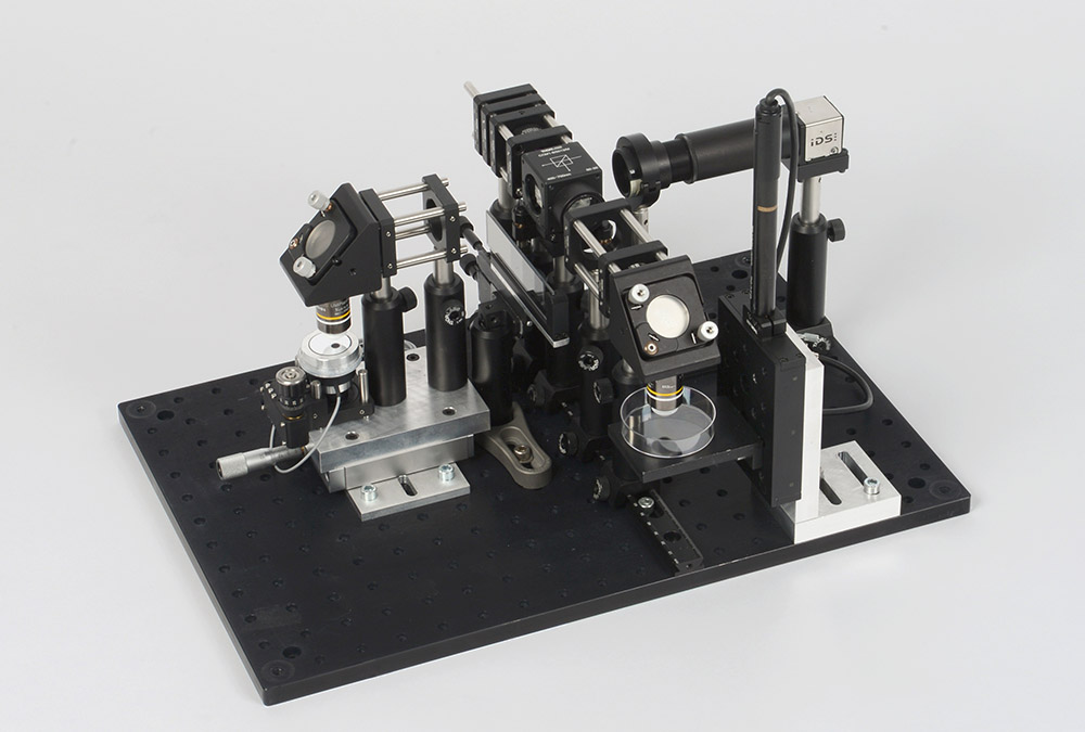
The non-invasive imaging of internal structures is of great importance for diagnostics in many medical disciplines. Especially in carcinoma diagnostics, however, there is a lack of high-resolution tomographic imaging techniques that can accelerate the analysis process of suspicious lesions. Measurement systems for optical coherence tomography (OCT) therefore offer great diagnostic added value due to their high resolution and measurement speed.
FF-OCT is an imaging technique that combines a high lateral resolution of light microscopy with the high axial resolution of conventional Spectral Domain or Swept Source OCTs. Like SD- and SS-OCT, FF-OCT is based on the principle of low coherent interferometry. In contrast to conventional OCT, the sample is illuminated by a microscope objective. The reflected light is detected and evaluated by an area scan camera. This results in high-resolution slice images from different depths of the sample. The slice images allow a fast tissue analysis on cellular level without the need of tissue manipulations.
Full-Field Optical Coherence Tomography (FF-OCT or Full-Field OCT) is ideally suited for the in vitro examination of tissue samples as well as for the in vivo examination of patient tissue. It has the potential, for example, to accelerate the process of preoperative and intraoperative evaluation of lesions and thus improve many aspects of diagnostic and therapeutic procedures. In preclinical studies, the Fraunhofer IPT deals with the development of FF-OCT systems, their application and their integration into imaging systems.
If the FF-OCT is designed as a tabletop system, volume images of tissue samples can be generated within a few minutes. When designed for in vivo use, for example by integration into an endoscope, cellular structures can also be displayed without tissue removal. This speeds up the workflow considerably and reduces the inconvenience and risks for patients. High-resolution tomographic tissue images are required not only for carcinoma diagnostics but also in other medical disciplines such as ophthalmology (retina and cornea diagnostics) and dermatology.
Our service
- Development of individual FF-OCT systems
- Implementation of application-specific signal and image processing algorithms
- Systems integration
- Implementation of preclinical feasibility studies

